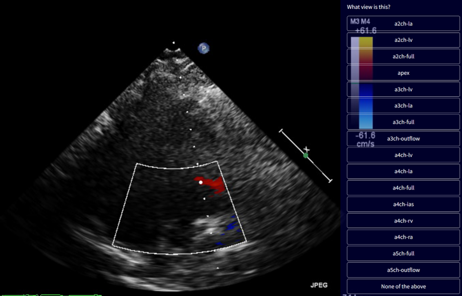
We are training an AI model for detailed classification of echocardiographic views, covering 47 distinct categories. Due to the large number of views, some challenging cases may arise. For instance, certain views, such as the apical 3-chamber and apical 4-chamber, can look very similar, making it difficult to determine the correct label. Please examine the image carefully to distinguish between these views. Additionally, if an image does not fit any of the available categories, select the "None of the above" option when labelling.
Watch a 1-minute demo video
Start Labelling
Description
Apical Views:
| View | Description |
|---|---|
| a2ch-la | Apical 2-chamber, focused on the left atrium |
| a2ch-lv | Apical 2-chamber, focused on the left ventricle |
| a2ch-full | Apical 2-chamber |
| apex | Any apical window, focused on the apex |
| a3ch-la | Apical 3-chamber, focused on the left atrium |
| a3ch-lv | Apical 3-chamber, focused on the left ventricle |
| a3ch-full | Apical 3-chamber |
| a3ch-outflow | Apical 3-chamber, focused on the aortic valve |
| a4ch-la | Apical 4-chamber, focused on the left atrium |
| a4ch-lv | Apical 4-chamber, focused on the left ventricle |
| a4ch-full | Apical 4-chamber |
| a4ch-ias | Apical 4-chamber, focused on the inter-atrial septum |
| a4ch-ra | Apical 4-chamber, focused on the right atrium |
| a4ch-rv | Apical 4-chamber, focused on the right ventricle |
| a5ch-full | Apical 5-chamber |
| a5ch-outflow | Apical 5-chamber, focused on the aortic valve |
Doppler Views:
| View | Description |
|---|---|
| doppler-ao-descending | Spectral Doppler of the descending aorta |
| doppler-mv | Spectral Doppler of the mitral valve |
| doppler-av | Spectral Doppler of the aortic valve |
| doppler-pv | Spectral Doppler of the pulmonary valve |
| doppler-tv | Spectral Doppler of the tricuspid valve |
| doppler-tissue-lateral | Tissue Doppler of the left ventricular lateral wall |
| doppler-tissue-septal | Tissue Doppler of the left ventricular septal wall |
| doppler-tissue-rv | Tissue Doppler of the right ventricular free wall |
PLAX Views:
| View | Description |
|---|---|
| plax-full-out | Parasternal long-axis, zoomed out |
| plax-full-lv | Parasternal long-axis, focused on the left ventricle |
| plax-full-la | Parasternal long-axis, focused on the left atrium |
| plax-full-rv-ao | Parasternal long-axis, focused on the right ventricle and aorta |
| plax-full-mv | Parasternal long-axis, centered on the mitral valve |
| plax-valves-av | Parasternal long-axis, focused on the aortic valve |
| plax-valves-mv | Parasternal long-axis, focused on the mitral valve |
| plax-tv | Parasternal inflow view including tricuspid valve |
PSAX Views:
| View | Description |
|---|---|
| psax-all | Parasternal short-axis, valve level, including all valves |
| psax-av | Parasternal short-axis, focused on aortic valve |
| psax-tv | Parasternal short-axis, focused on tricuspid valve |
| psax-pv | Parasternal short-axis, focused on pulmonary valve |
| psax-lv-base | Parasternal short-axis, left ventricle base level |
| psax-lv-mid | Parasternal short-axis, left ventricle mid-level |
| psax-lv-apex | Parasternal short-axis, left ventricle apex level |
M-mode Views:
| View | Description |
|---|---|
| mmode-a4ch-rv | M-mode for measuring TAPSE |
| mmode-ivc | M-mode of the inferior vena canva |
| mmode-plax-mitral | M-mode of the mitral valve in the parasternal long-axis |
| mmode-plax-av | M-mode of the aortic valve in the parasternal long-axis |
| mmode-plax-lv | M-mode of the left ventricle in the parasternal long-axis |
Subcostal/Suprasternal Views:
| View | Description |
|---|---|
| subcostal-heart | Subcostal window, focused on the heart |
| subcostal-ivc | Subcostal window, focused on the inferior vena canva |
| suprasternal | Suprasternal view |