Project Introduction
Quantification of the left ventricle shape is crucial in evaluating cardiac function from
2D echocardiographic images. This study investigates the applicability of established loss
functions when optimising the U-Net model for 2D echocardiographic left ventricular segmentation.
Our results indicate loss functions are a significant component for optimal left
ventricle volume measurements when established segmentation metrics could be imperceptible.
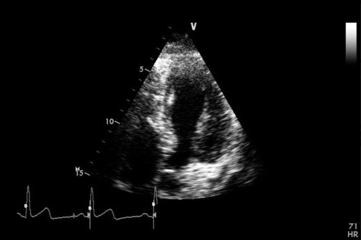
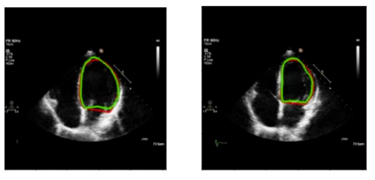
Fig. 1. An example from the dataset
Dataset
1224 Videos of the apical 4-chamber echocardiographic view, acquired between 2015 and 2016,
were extracted from Imperial College Healthcare NHS Trust’s echocardiogram database.
The acquisition of the images was performed by experienced echocardiographers and according to standard protocols,
using ultrasound equipment from GE and Philips manufacturers.
Ethical approval was obtained from the Health Regulatory Agency (Integrated Research Application System identifier 243023).
From these videos, a total of 2600 images were sampled from different points in the cardiac cycle. Each image underwent labelling
by one individual from a pool of experts using our in-house online labelling platform
(https://unityimaging.net). This dataset was used for model developments (i.e., training and validation).
The testing comprised 100 videos, from consecutive studies conducted over 3 working
days in 2019, at least 3 years away from the date of collection for the model development dataset.
Our previous deep neural network (Lane et al., 2021) was used to identify the
end-diastolic and end-systolic frames of the 100 videos. These selected frames were used
for the human expert annotations. Each of the 200 resulting images was then labelled
by 11 experts, using the platform. The concensus of the expert was finally used as the
ground-truth in the testing dataset.
Method
U-Net was implemented in TensorFlow and was trained on an Nvidia RTX3090 GPU.
The loss functions chosen for experimentation are common for image segmentation tasks
and were selected from three categories:
• Distribution-based loss: Binary cross entropy (BCE) loss
• Region-based loss: Dice loss, Tvsersky loss, Focal Tvsersky loss
• Compound loss: BCE+Dice
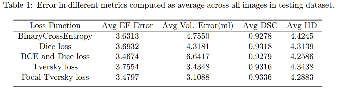
After training the model for different loss functions, each model was evaluated using the
established evaluation metrics for segmentation tasks (i.e., Dice Coefficient and Hausdorff
Distance) and domain specific metrics (i.e., volume and EF measurements) by averaging the
error across all images in the testing dataset. The volume was computed using the Simpson’s method.
The average Volume Error is calculated using Cartesian pixel coordinates
by computing the difference between the volume of the ground-truth and the predicted
endocardial border.
EF was estimated by dividing the stroke volume (i.e., the difference between end-diastolic
and end-systolic volumes) by the end-diastolic volume.
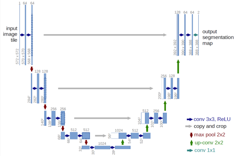
Fig. 2. U-Net Model
Experiment Results and Discussion