Project Description
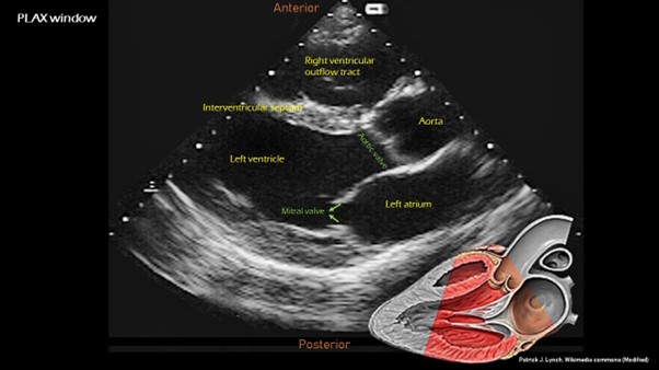
Visualizing the mitral valve (MV) in its entirety in PLAX images is essential for comprehensive cardiac evaluation. This project aims to create a standardized, open-access dataset of PLAX images focusing on full MV views, annotated by experts.
Project Goals
- Develop a Reference Dataset: Compile a comprehensive dataset of PLAX images highlighting full MV views, annotated by experienced echocardiographers.
- Standardize Full MV Assessment: Establish objective criteria for evaluating the quality of full MV views in PLAX images.
- Empower AI Development: Provide a high-quality dataset to train and validate AI algorithms for detecting and enhancing full MV views.
- Improve Image Acquisition: Develop training materials and guidelines to help sonographers optimize image acquisition for clear full MV views.
- Enhance Diagnostic Accuracy: Improve the reliability of PLAX echocardiographic interpretations for better patient care.
Your Role
- Review PLAX Echocardiographic Images: You will be presented with a series of PLAX echocardiographic images.
- Assess Full MV Views: Use the provided guidelines and your expertise to assess the quality of full MV views in each image.
- Rank Images: Rank the images based on the clarity and completeness of the full MV views.
- Provide Feedback (Optional): Share your insights and suggestions for improving the annotation process or the assessment criteria.
How to Participate
- Create an Account: Register on the UnityImaging.net labelling platform.
- Access the Project: Join the "PLAX Full-MV MV Assessment Project."
- Review Guidelines: Familiarize yourself with the detailed annotation instructions and criteria for assessing full MV views.
- Start Annotating: Begin reviewing and ranking PLAX images based on the quality of full MV views.
- Provide Feedback (Optional): Share your insights to help refine the process and improve the dataset.
Ranking Criteria
- Mitral Valve Visualization: Assess how well the mitral valve is visualized in its entirety.
- Overall Image Quality: Rate the overall clarity and diagnostic quality of the image, considering factors like contrast and resolution.
Who Should Participate?
- Echocardiographers: Your expertise in image interpretation is crucial for accurate assessment.
- Cardiologists: Your clinical knowledge can help understand the impact of full MV views on diagnosis
- Sonographers: Your experience in image acquisition can provide valuable insights into optimizing image quality.